
Revolutionary SPIFFI Microscopy Technique Produces Super-Resolution Cellular Videos from a Single Exposure

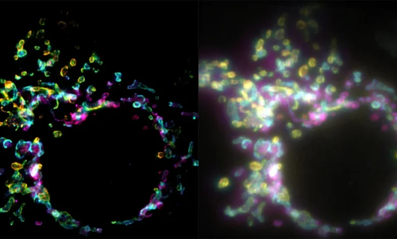
In a breakthrough that promises to reshape the landscape of cellular biology, researchers have unveiled a novel fluorescence microscopy technique capable of generating super-resolution images from a single exposure. Developed by a team at the Laboratory of Nanoscale Biology (LBEN) within EPFL’s School of Engineering, the method is known as SPIFFI—an acronym for spatial polarization-induced fluorescence fluctuation imaging. Published in the journal Nature, this development overcomes a fundamental bottleneck that has constrained optical microscopy for decades: the inability to capture high-resolution imagery of fast-moving biological processes inside living cells.
Traditional optical microscopy has long been bound by the diffraction limit of light, a physical constraint that prevents conventional microscopes from distinguishing structural details smaller than approximately 200 to 300 nanometers. While the advent of super-resolution fluorescence microscopy in the late 20th and early 21st centuries—culminating in the 2014 Nobel Prize in Chemistry—shattered this barrier, it introduced a severe secondary limitation. These legacy super-resolution methods typically rely on temporal information, requiring the capture of hundreds or thousands of sequential frames to reconstruct a single, sharp image.
While this multi-frame approach works remarkably well for fixed, static samples, it fundamentally fails when applied to living cells. Cellular environments are dynamic, fluid, and in a constant state of motion; organelles shift, membranes fuse, and proteins migrate on millisecond timescales. Attempting to stitch together hundreds of frames to form a single image of a living specimen is akin to taking a long-exposure photograph of a bustling highway—the result is an unusable blur.
Addressing the Limitations of Temporal Imaging
The development of SPIFFI addresses this long-standing dilemma by shifting the analytical paradigm from time to space. Instead of aggregating data across numerous temporal frames, SPIFFI extracts high-resolution spatial details instantaneously from a single snapshot.
The core mechanics of the technique hinge on the photophysical properties of fluorescent molecules, known as fluorophores. When these molecules are excited by light and emit their own fluorescence, the emitted light waves are not uniform. Instead, they oscillate preferentially in specific directions, a phenomenon dictated by the precise spatial orientation of the molecule at the moment of emission.
Wei Guo, an LBEN PhD student and the primary author of the research paper, elucidates the fundamental shift in methodology. Previous approaches utilized temporal information to resolve spatial limitations, a strategy fundamentally incompatible with the fluid nature of living cellular architecture. By contrast, SPIFFI leverages the inherent polarization of light. The optical system is engineered to capture and split the fluorescent light emitted by the sample into four distinct polarization-sensitive channels. By comparing the subtle variations and fluctuations across these four parallel channels, the system mathematically recovers structural details that would otherwise remain hidden under a standard widefield lens.
Experimental Validation and Performance Metrics
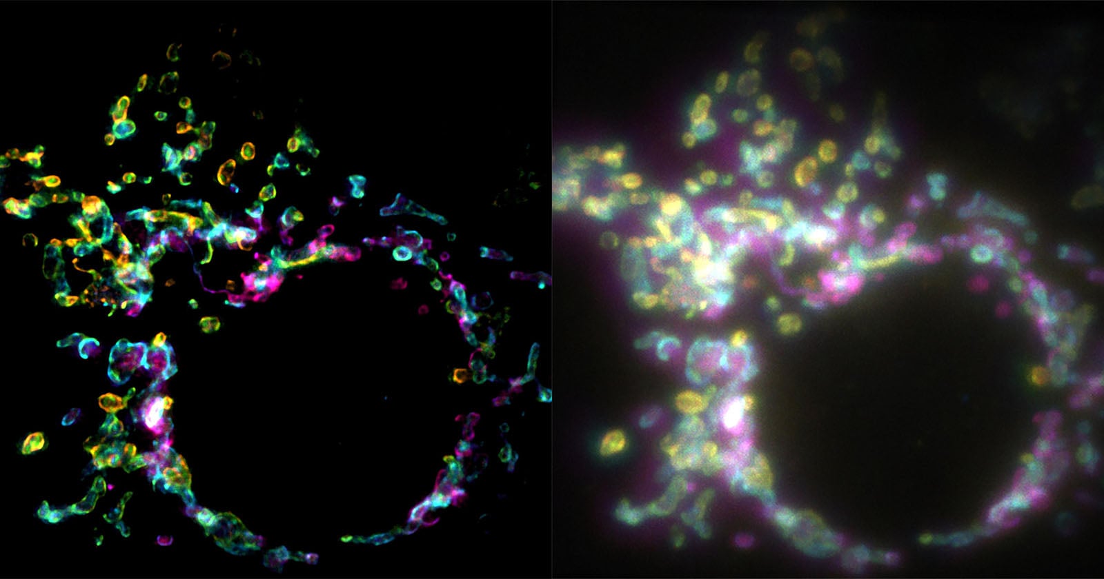
In rigorous laboratory testing, the research team—which includes Wei Guo, Lely Feletti, and Aleksandra Radenovic—demonstrated that SPIFFI effectively doubles image resolution within a single exposure. The system successfully resolved delicate cellular structures approximately 160 to 170 nanometers in size. More importantly, this capability was deployed to sharply image highly dynamic, transient cellular events, including the physical splitting (cell division) and fusion of membranes.
Radenovic, heading the LBEN team, notes that the implications extend far beyond simple snapshot clarity. Because every individual frame captured by the SPIFFI system is inherently super-resolved, researchers are no longer restricted to producing static pictures of dead cells. They can now generate true super-resolution videos of live biological processes unfolding in real time.
Furthermore, the research team discovered that SPIFFI’s output is not mutually exclusive with existing technology. By seamlessly integrating SPIFFI imagery with established fluctuation-based post-processing algorithms, the researchers achieved an even more refined resolution of approximately 80 nanometers, pushing the boundaries of what is optically achievable with standard fluorophores.
Hardware Adaptability and Practical Integration
Beyond its theoretical and analytical breakthroughs, SPIFFI offers a significant practical advantage for academic and commercial laboratories: hardware accessibility. Many cutting-edge super-resolution modalities require entirely custom, highly expensive microscope builds that occupy dedicated laboratory spaces and demand specialized maintenance.
In contrast, the optical hardware configuration required for SPIFFI is designed to be retrofitted onto existing fluorescence microscopes. By modifying the optical path to split and analyze the polarization channels, laboratories can upgrade their current hardware suites rather than investing in entirely new imaging platforms. Looking forward, the EPFL research team is actively engineering more compact iterations of the SPIFFI hardware, a development intended to streamline usability and accelerate adoption across broader scientific communities.
Broader Implications for Biological and Medical Research
The ability to observe live intracellular mechanics at unprecedented resolutions opens up vast avenues of inquiry across multiple scientific disciplines. Virology, oncology, and neurobiology stand to benefit immensely from a tool capable of tracking viral entry, intracellular drug delivery, and synaptic vesicle dynamics without the motion-blur artifacts characteristic of legacy systems.
As researchers worldwide gain access to SPIFFI-compatible configurations, the scientific community anticipates a paradigm shift in how dynamic cellular pathology is studied. By transforming what was once a multi-frame composite into an instantaneous, high-definition reality, SPIFFI marks a defining milestone in the evolution of optical microscopy.